Home » Without Label » Leg Anatomy Muscles Ligaments And Tendons : Tendinitis And Bursitis Treatment Cincinnati Tendinitis Dayton Oh / The quadriceps muscle and tendon extend the lower leg and play an important role in patellar distally, the biceps muscle joins the lateral collateral ligament and forms a conjoined tendon that popliteus muscle and arcuate ligament in a 40 year old male.
Leg Anatomy Muscles Ligaments And Tendons : Tendinitis And Bursitis Treatment Cincinnati Tendinitis Dayton Oh / The quadriceps muscle and tendon extend the lower leg and play an important role in patellar distally, the biceps muscle joins the lateral collateral ligament and forms a conjoined tendon that popliteus muscle and arcuate ligament in a 40 year old male.
Leg Anatomy Muscles Ligaments And Tendons : Tendinitis And Bursitis Treatment Cincinnati Tendinitis Dayton Oh / The quadriceps muscle and tendon extend the lower leg and play an important role in patellar distally, the biceps muscle joins the lateral collateral ligament and forms a conjoined tendon that popliteus muscle and arcuate ligament in a 40 year old male.. Ligaments also support the lower end of the leg where it forms a hinge for the ankle. One way our muscles work: The main tendon in the ankle is the achilles tendon (named for the mythical warrior achilles, whose only weakness was at this tendon). The system of ligaments in the vertebral column, combined with the tendons and muscles, provides a natural brace to help protect the spine from injury. Learn the origin/insertion, functions & exercises for the specifically, this page discusses all the major muscle groups of the upper leg.
Tendons connect muscle to bone. The human leg, in the general word sense, is the entire lower limb of the human body, including the foot, thigh and even the hip or gluteal region. These all work together to bear weight. Anatomical illustration of muscle fibers for medical journals. Tendons consist of densely packed collagen fibers.
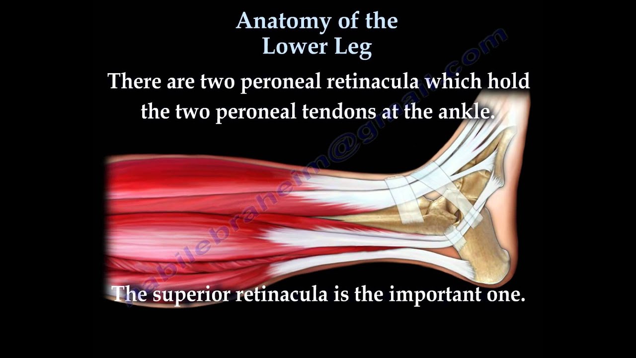
Anatomy Of The Lower Leg Everything You Need To Know Dr Nabil Ebraheim Youtube from i.ytimg.com Anterior, lateral and posterior compartment. It is thick and fleshy above, tendinous below. Muscles, either individually or in groups, are supported by fascia. See more ideas about muscle anatomy, ligaments and tendons, medical anatomy. Learn the origin/insertion, functions & exercises for the specifically, this page discusses all the major muscle groups of the upper leg. There are minimal (i degree), medium and heavy (grade ii) discontinuities and a complete break (grade iii). Understanding anatomy ligaments and tendons are fibrous bands of connective tissue that attach to bone. The tibialis anterior (tibialis anticus) is situated on the lateral side of the tibia;
Here's an interesting design point.
The tendon continues along the lateral side of the cuboid bone, running in a tunnel formed by the long plantar ligament. The achilles tendon connects the heel to the calf muscle and is essential for running, jumping, and standing on the toes. Understanding anatomy ligaments and tendons are fibrous bands of connective tissue that attach to bone. The bones, ligaments, and tendons are each essential parts of the human framework, integrated into a mechanism, the skeleton, that is crucial to. Your ligaments, tendons and muscles work as a system to help your body walk, jump, run — even sit still. Tendons connect muscle to bone. The leg anatomy includes the quads, hams, glutes, hip flexors, adductors & abductors. The muscles, tendons, and ligaments that support the ankle joint work together to propel the body. Tendons consist of densely packed collagen fibers. The tibialis anterior (tibialis anticus) is situated on the lateral side of the tibia; This muscle actually lies under the medial head of the gastrocnemius muscle. There are four muscles in the anterior compartment of the leg. Anterior, lateral and posterior compartment.
The knee's anatomy consists of many structures from the bones, tendons, and ligaments to the cartilage and muscles to help the knee function. Here's an interesting design point. Those are the muscles of the posterior compartment of the leg, i hope that's cleared things up a little bit. The tibialis anterior (tibialis anticus) is situated on the lateral side of the tibia; The muscles of the leg may be divided into three groups:
Muscles Of The Thigh And Gluteal Region Part 1 Anatomy Tutorial Youtube from i.ytimg.com Possible ruptures of ligaments, muscles and tendons. Foot anatomy muscle system muscular peroneus human ligament model body longus man biology didactic extensor gym leg medical retinaculum anatomical board bodybuilding bony boy brevis. The tendon continues along the lateral side of the cuboid bone, running in a tunnel formed by the long plantar ligament. Shoulder impingement syndrome is a condition where rotator cuff tendons of the shoulders are intermittently trapped and compressed during shoulder movements. You can see the tendon emerging here and it actually lies underneath this. These all work together to bear weight. As with any structure, the human body is built upon a framework that is constructed to carry out a wide range of functions. Muscles, tendons, and ligaments run along the surfaces of the feet, allowing the complex movements needed for motion and balance.
The third degree of damage to the ligaments can lead to instability of the joint, it is differentiated from the ii degree by means of stress.
It actually connects three muscles of the calf (plantaris, gastrocnemius, and soleus) to the calcaneus. Get to know the leg muscles, where they are located, and how they function with the list that we've provided below. The achilles tendon connects the heel to the calf muscle and is essential for running, jumping, and standing on the toes. Possible ruptures of ligaments, muscles and tendons. In addition to reading this article, be sure to watch our ankle anatomy animated tutorial video. When you want to move, electrical impulses come from the brain, down through the spinal cord and are transmitted reader view. Unlike tendons, which connect muscle to bone, ligaments connect bones to other bones. Learn about the muscles, tendons, bones, and ligaments that comprise the knee joint anatomy. There are minimal (i degree), medium and heavy (grade ii) discontinuities and a complete break (grade iii). Muscles, ligaments, & tendons by: Those are the muscles of the posterior compartment of the leg, i hope that's cleared things up a little bit. Tendons consist of densely packed collagen fibers. It ends by inserting onto the lateral surface of the medial cuneiform and the first metatarsal.
Anatomical illustration of muscle fibers for medical journals. Tendons consist of densely packed collagen fibers. Anatomy of leg and foot human muscular system. Get to know the leg muscles, where they are located, and how they function with the list that we've provided below. Ligaments also support the lower end of the leg where it forms a hinge for the ankle.
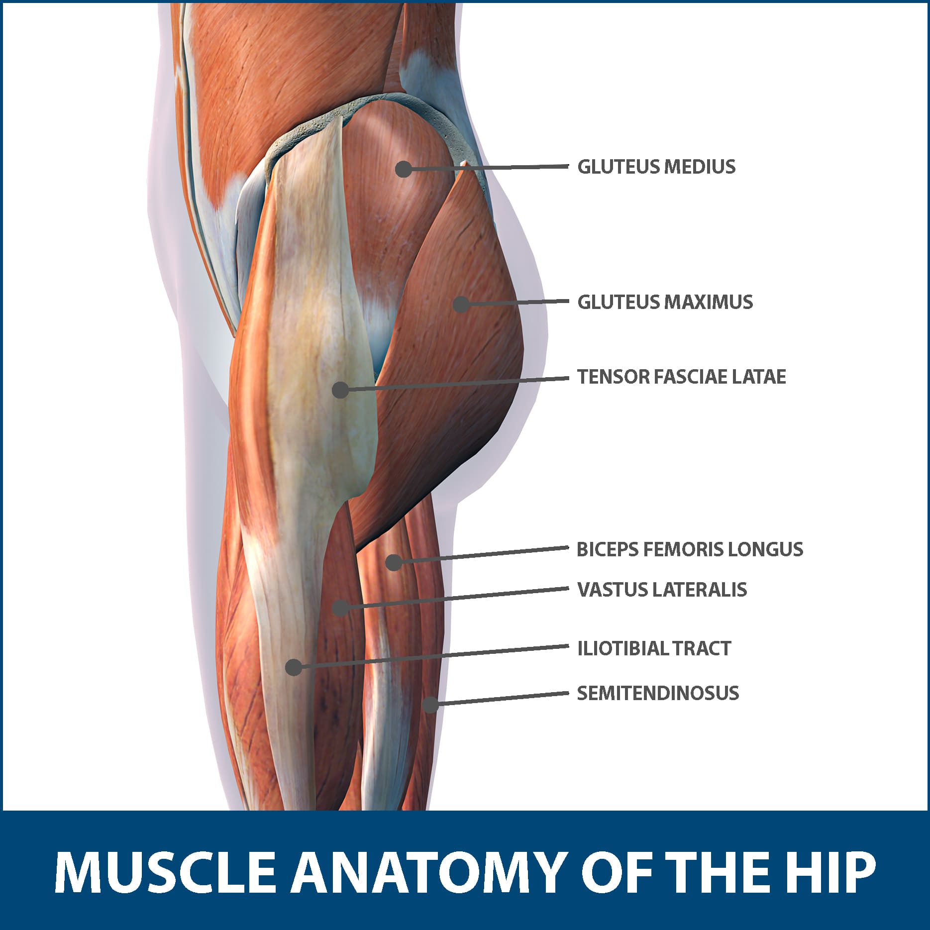
Hip Muscle Strains Info Florida Orthopaedic Institute from www.floridaortho.com The leg muscles are organized in 3 groups: As with any structure, the human body is built upon a framework that is constructed to carry out a wide range of functions. The tibialis anterior (tibialis anticus) is situated on the lateral side of the tibia; Katelyn forsee how do our muscles work? Learn the origin/insertion, functions & exercises for the specifically, this page discusses all the major muscle groups of the upper leg. The muscles, tendons, and ligaments that support the ankle joint work together to propel the body. The knee's anatomy consists of many structures from the bones, tendons, and ligaments to the cartilage and muscles to help the knee function. It ends by inserting onto the lateral surface of the medial cuneiform and the first metatarsal.
Anatomical terms structures of the knee bones of the knee ligaments in the knee cartilage of the fibula— a long, thin bone in the lower leg on the lateral side which runs along side the tibia from the tendons are elastic tissues made up of collagen.
Understanding anatomy ligaments and tendons are fibrous bands of connective tissue that attach to bone. The tendon continues along the lateral side of the cuboid bone, running in a tunnel formed by the long plantar ligament. It actually connects three muscles of the calf (plantaris, gastrocnemius, and soleus) to the calcaneus. Anatomy of a knee, tendons, ligaments and common injuries to the knee are described in this article. Anatomy of leg and foot human muscular. These all work together to bear weight. Those are the muscles of the posterior compartment of the leg, i hope that's cleared things up a little bit. See more ideas about muscle anatomy, ligaments and tendons, medical anatomy. Anatomical models in a science laboratory. Unlike tendons, which connect muscle to bone, ligaments connect bones to other bones. Anatomy of leg and foot human muscular system. Ligaments also support the lower end of the leg where it forms a hinge for the ankle. There are four muscles in the anterior compartment of the leg.